


3D Cell Painting Reveals Cancer Drug Differences Missed in 2D Screens
Discover how 3D Cell Painting and AI-driven imaging reveal cancer drug responses missed by traditional 2D screens, advancing precision oncology.
Cancer drug development has traditionally relied on two-dimensional (2D) cell cultures for initial screening and phenotypic analysis. While comprehensive and cost-effective, 2D models often fail to replicate the complex cellular architectures and microenvironmental cues present in vivo, leading to gaps in understanding drug efficacy and mechanism of action. Recent advancements in three-dimensional (3D) cell culture combined with high-content imaging modalities, such as 3D Cell Painting, have begun to address these limitations, revealing subtle but critical differences in cancer drug responses that 2D assays may overlook.
Understanding 3D Cell Painting
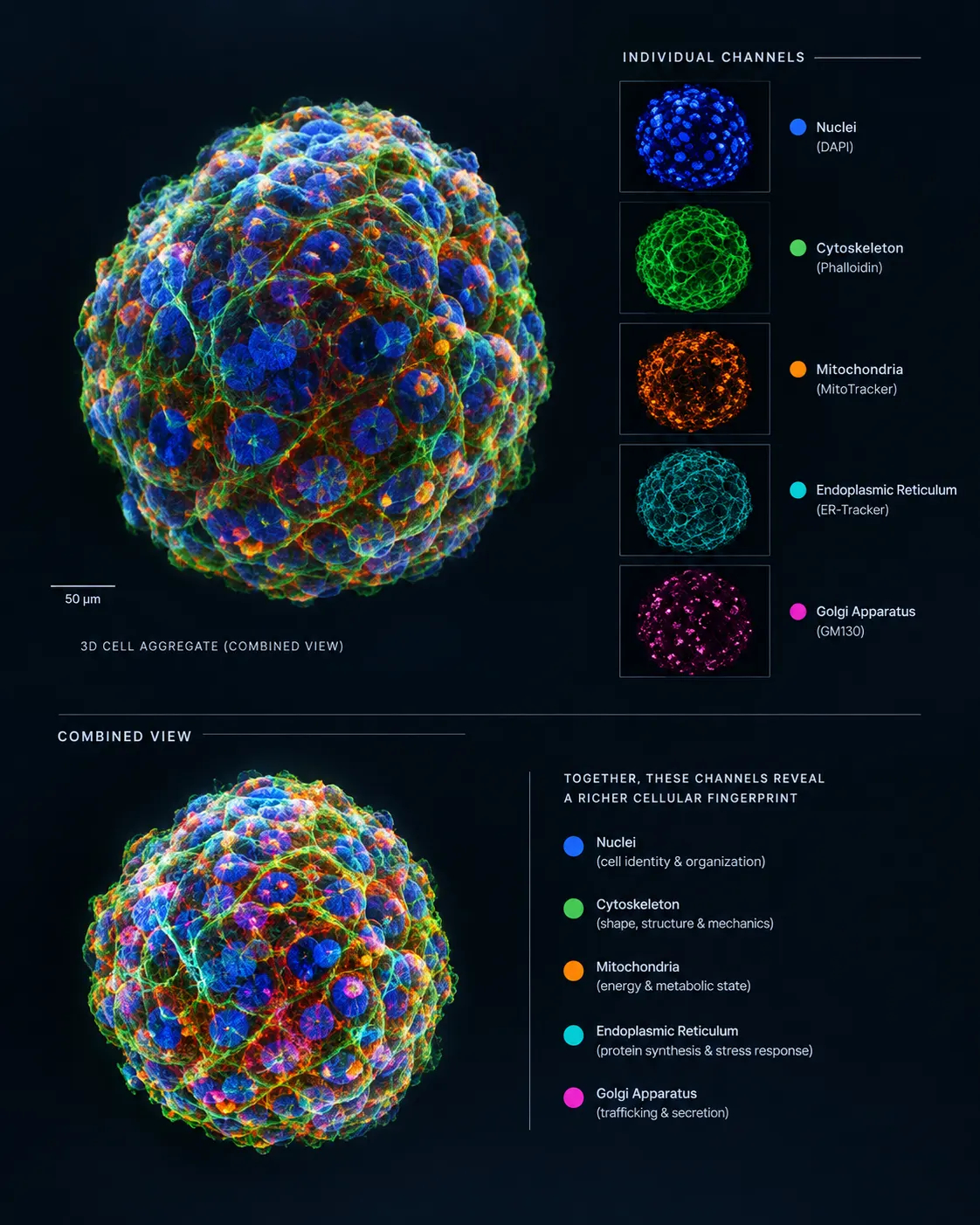
Cell Painting is a multiplexed, high-content imaging technique that employs a palette of fluorescent dyes to label and visualize distinct cellular components including nuclei, cytoskeleton, mitochondria, endoplasmic reticulum and Golgi apparatus. Combined with automated image analysis and machine learning, this method generates rich morphological fingerprints that quantify phenotypic alterations induced by chemical or genetic perturbations.
Extending Cell Painting to 3D culture systems involves growing cancer cells in spheroids or organoids that better mimic tissue complexity, cell–cell and cell–matrix interactions and gradients of nutrients and oxygen. Imaging these 3D cultures in volumetric space provides a multidimensional phenotypic readout capturing spatial heterogeneity and cellular organization absent from planar 2D monolayers.
Why 3D Models Reveal More About Drug Effects
Cancer is inherently a three-dimensional disease. Tumor cells interact dynamically with their microenvironment, including stromal cells and extracellular matrix, influencing drug penetration, metabolism and resistance mechanisms. In 2D cultures, the absence of these factors can mask critical phenotypic responses to oncology therapeutics.
Recent studies have demonstrated that certain anticancer agents produce distinct morphological signatures in 3D spheroids compared to 2D cultures. For instance, drugs targeting cytoskeletal elements or mitochondrial function may induce alterations in cellular architecture or metabolic heterogeneity that only become apparent when cells assemble into 3D structures. Additionally, drug efficacy and toxicity profiles can differ markedly, highlighting potential false negatives or positives in purely 2D-based screens.
Examples of Drug Differences Uncovered by 3D Cell Painting
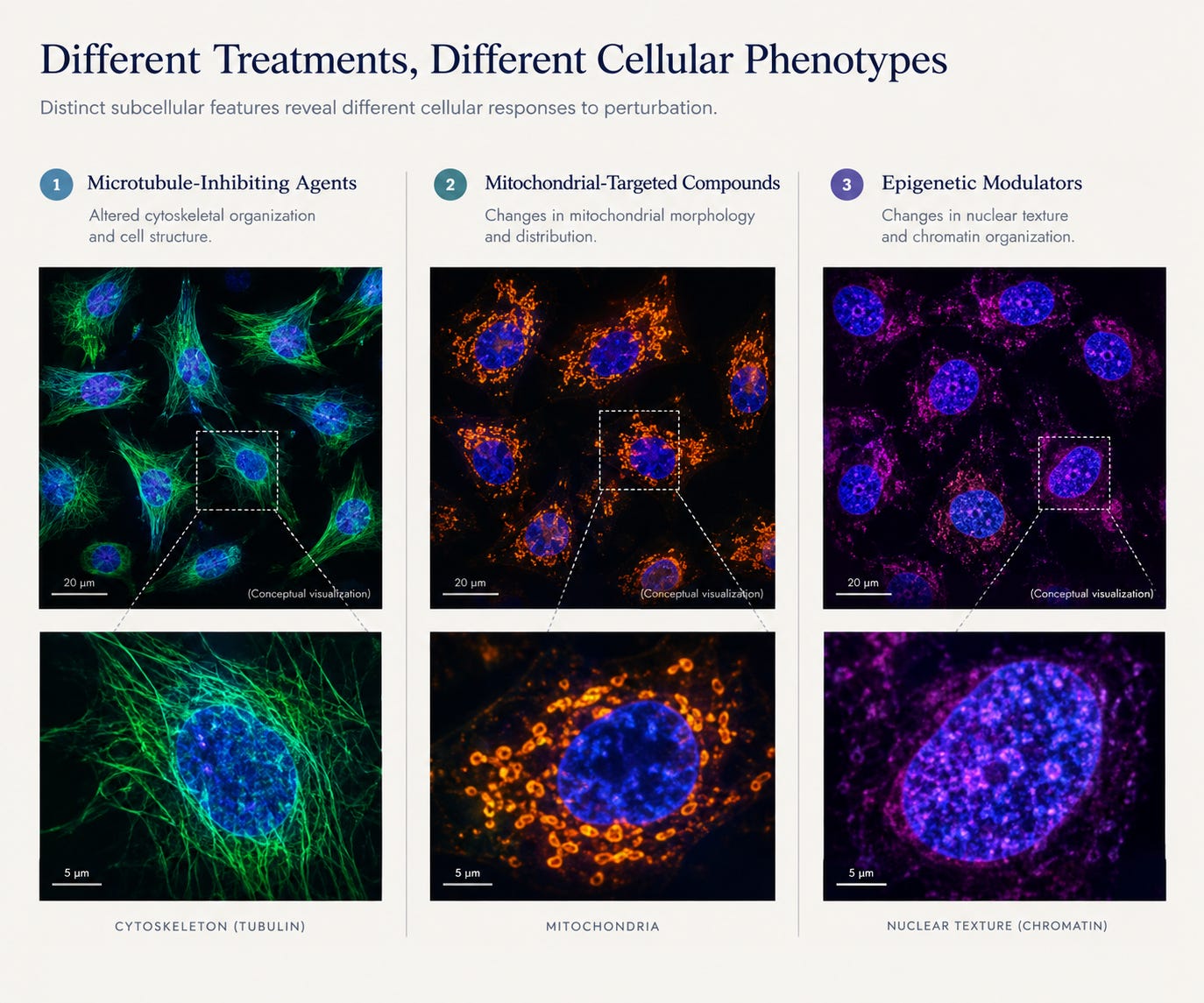
- Microtubule-Inhibiting Agents: Agents such as paclitaxel can display altered microtubule stabilization dynamics in 3D spheroids, changing nuclear morphology and mitotic index more clearly than in 2D. These differences impact interpretations of drug potency and mechanism.
- Mitochondrial-Targeted Compounds: Metabolic gradients in 3D cultures accentuate mitochondrial dysfunction induced by specific drugs, leading to distinct mitochondrial morphology and membrane potential changes that 2D approaches often miss.
- Epigenetic Modulators: Agents modulating chromatin remodelers may induce differential nuclear texture and chromatin condensation in 3D contexts, reflecting more accurately their role in gene expression plasticity within tumors.
Integrating AI in 3D Cell Painting Enhances Drug Discovery Insights
Given the multidimensionality and data richness of 3D Cell Painting assays, advanced AI-driven computational approaches are essential for effective analysis. Machine learning models can integrate morphological features across hundreds of cellular parameters, facilitating the classification of subtle phenotypes related to mechanism of action, off-target effects or synergistic drug combinations.
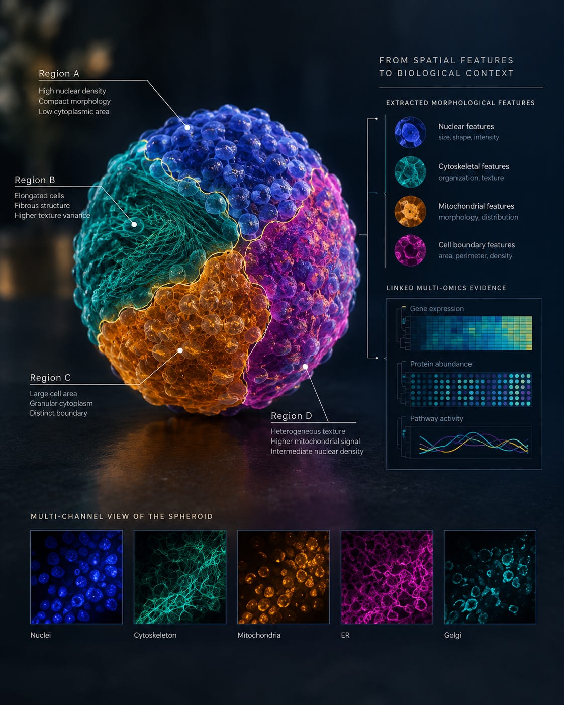
Medvolt leverages deep learning algorithms and data integration frameworks to refine phenotypic signatures derived from 3D Cell Painting, correlating morphological fingerprints with multi-omics profiles and clinical outcomes. This systems biology perspective enables a more precise deconvolution of drug responses, supporting lead optimization and patient stratification efforts.
The Value of 3D Cell Painting in Personalized Oncology
Moving beyond drug discovery, 3D Cell Painting allows the analysis of patient-derived tumor cells cultured as organoids, capturing tumor heterogeneity and microenvironment interactions. Morphological profiling combined with AI can identify patient-specific drug sensitivities, informing personalized treatment regimens.
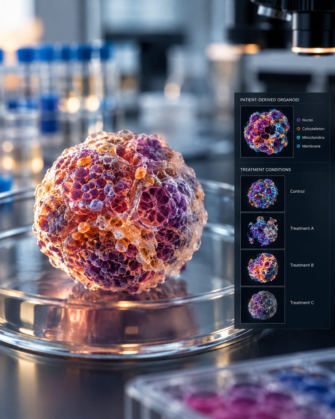
Moreover, 3D profiling reveals mechanisms of resistance evolving within the tumor microenvironment, guiding the design of combination therapies that address emergent phenotypes not evident in 2D models.
Challenges and Future Directions
While promising, 3D Cell Painting introduces complexities related to imaging depth, signal attenuation and increased computational requirements. Advances in light-sheet microscopy and volumetric image processing algorithms continue to enhance data quality and throughput.
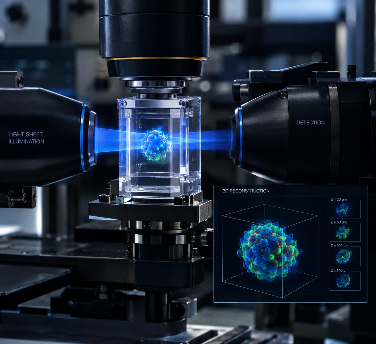
Further integration with computational chemistry techniques such as free energy perturbation (FEP) simulations and fragment-based drug design can link morphological phenotypes with molecular interactions, closing the loop between cell biology and medicinal chemistry.
Conclusion
The shift from 2D to 3D Cell Painting represents a significant evolution in phenotypic drug screening, unveiling cancer drug effects obscured in traditional models. By capturing cellular architecture and microenvironmental context, 3D profiling deepens mechanistic understanding and improves predictive accuracy in oncology drug discovery.
Medvolt’s commitment to AI-driven analysis and multi-omics integration positions it at the forefront of leveraging 3D Cell Painting data to accelerate novel cancer therapeutics development and precision medicine applications. This approach exemplifies how combining innovative biology with computational power can yield a more nuanced and clinically relevant picture of drug action.

