


Automated MRI Pipeline Standardises Preclinical Stroke Damage Measurement
Explore how automated MRI pipelines and AI-driven image analysis improve stroke lesion measurement, reproducibility and preclinical neurovascular research.
Stroke remains a leading cause of disability and mortality worldwide, prompting vigorous research efforts toward understanding its pathophysiology and developing effective interventions. Preclinical models, particularly rodent ischemic stroke models, are central to this endeavor, enabling the evaluation of neuroprotective strategies and therapeutic candidates before entering clinical trials. However, precise and reproducible quantification of stroke-induced brain damage has historically presented significant challenges. Variability in manual lesion delineation and imaging protocols hinders consistent cross-study comparisons and increases the risk of experimental bias. In this context, the advent of automated MRI pipelines represents a significant advancement in preclinical stroke research, enabling standardised, high-throughput, and objective measurement of cerebral infarcts.
Challenges in Preclinical Stroke Imaging
Magnetic resonance imaging (MRI) has become a gold standard modality for assessing cerebral ischemic injury in animal models. Its non-invasive nature allows longitudinal monitoring of lesion evolution, providing critical insights into injury dynamics and therapeutic responses. Techniques such as T2-weighted imaging (T2WI) and diffusion-weighted imaging (DWI) are commonly employed to visualize infarcted regions and peri-infarct zones. Nevertheless, the quantification of stroke damage from these imaging data largely depends on manual or semi-automated segmentation approaches.
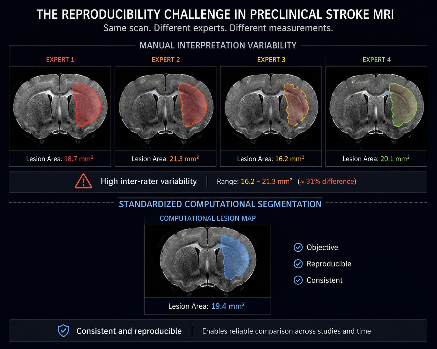
Manual tracing of infarcts is highly operator-dependent and can introduce subjective bias, affecting the reproducibility and reliability of lesion volume measurements. Moreover, inconsistencies in delineation criteria across laboratories and studies complicate data harmonization. The time-consuming nature of such manual workflows also limits scalability, which is particularly problematic given the growing scale of preclinical studies evaluating multiple interventions and experimental conditions.
The Role of Automated MRI Pipelines
Automated MRI pipelines address these limitations by integrating machine learning algorithms and image processing techniques to standardise stroke lesion segmentation and quantification. These pipelines systematically process raw imaging data through a series of computational steps including noise reduction, image registration, tissue classification, and lesion detection—yielding objective and reproducible infarct maps.
Key features of an automated pipeline typically include:
- Preprocessing: Correction for motion artifacts, intensity normalization, and alignment of images to a standard brain atlas facilitate consistent analysis across subjects.
- Segmentation: Utilising supervised or unsupervised learning models, the pipeline differentiates infarcted tissue from healthy parenchyma based on signal intensity and morphological criteria.
- Quantification: Calculated lesion volumes and spatial distribution metrics are generated automatically, providing quantitative endpoints for downstream statistical analyses.
Such automation reduces human error, accelerates data throughput, and enables the inclusion of larger cohorts, enhancing statistical power and experimental rigour.
Integrating AI-driven Technologies for Enhanced Precision
Recent advancements in artificial intelligence (AI), particularly deep learning, have significantly improved segmentation accuracy in complex neuroimaging datasets. Convolutional neural networks (CNNs) trained on annotated preclinical MRI scans can learn intricate features of ischemic lesions, outperforming conventional threshold-based or atlas-driven methods.
By incorporating AI models, automated MRI pipelines can adapt to heterogeneous data arising from variable imaging protocols or different stroke induction models. This flexibility is critical for multi-center studies aiming for harmonized stroke damage assessment. Furthermore, AI algorithms can facilitate multimodal data integration—combining structural MRI with perfusion or functional imaging modalities—providing richer phenotypic descriptions of ischemic injury.
Implications for Drug Discovery and Neurovascular Research
Quantitative, standardised measurement of stroke lesions is indispensable for evaluating candidate therapeutics targeting neuroprotection, neuroinflammation, and recovery mechanisms. Precision in infarct volume assessment directly influences dose-response interpretations, efficacy comparisons, and identification of biomarkers predicting treatment outcomes.
Automated MRI pipelines contribute to improving reproducibility and transparency of preclinical data—factors increasingly emphasized by funding agencies, regulatory bodies, and journals. Enhanced data quality accelerates the translation of promising therapies into clinical settings by providing robust preclinical endpoints.
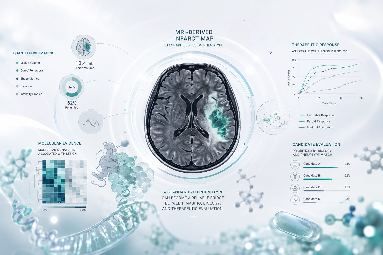
Moreover, automated analytics frameworks can be integrated with AI-driven drug discovery platforms. By linking lesion phenotypes with multi-omics and molecular modeling data, such integration opens new avenues for exploring mechanistic insights and identifying novel therapeutic targets. Computational workflows thereby support iterative cycles of hypothesis generation and experimental validation, amplifying research efficiency.
Medvolt’s Contribution and Vision
Medvolt leverages its expertise in AI and computational biology to develop and refine automated imaging pipelines tailored for preclinical neuroscience applications. By combining advanced image analysis algorithms with scalable cloud-based infrastructure, Medvolt enables high-throughput, reproducible stroke damage quantification.
Their platforms emphasize interoperability with diverse imaging modalities and experimental designs, facilitating collaborative research efforts and multi-center data pooling. Medvolt’s commitment to transparency includes rigorous validation protocols and open benchmarking against manual annotations, ensuring scientific credibility and adoption within the neurovascular research community.
In addition, Medvolt’s integrated AI-driven drug discovery environment synergistically connects imaging-derived phenotypes with molecular simulation and enzyme engineering tools. This holistic approach accelerates target identification, drug candidate optimization, and preclinical validation—addressing critical bottlenecks in stroke therapeutic development.
Conclusion
Automated MRI pipelines represent a pivotal advancement in preclinical stroke research, offering standardised, objective, and scalable measurement of cerebral infarcts. By reducing operator bias and enabling large-scale analyses, these pipelines enhance data reproducibility and interpretability, crucial for reliable therapeutic screening.
The integration of AI methods further refines lesion detection and expands analytical capabilities, empowering precision neuroscience investigations. Platforms like those developed by Medvolt bridge imaging biomarkers with computational drug discovery, fostering a synergistic environment for innovation in neurotherapeutics.
As the field continues to evolve, adoption of automated, AI-enabled imaging workflows will be fundamental to overcoming existing limitations in stroke modeling and expediting the translation of effective treatments to clinical practice.

